在临床前实验中,大小鼠常作为研究对象来模拟人类世界中的真实情况,以小鼠为例,在短短的2-3年中,大小鼠以比人更快的速度走完生命的每个阶段。

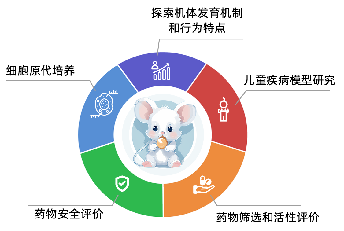
尤其在1月龄以前,小鼠年龄变化速度是人类的150倍[1]。0-21日龄之间的大小鼠对应人类婴儿至青春早期,这期间乳鼠的外观变化甚至以天为单位。 在实验应用上,乳鼠可以应用于以下方面:

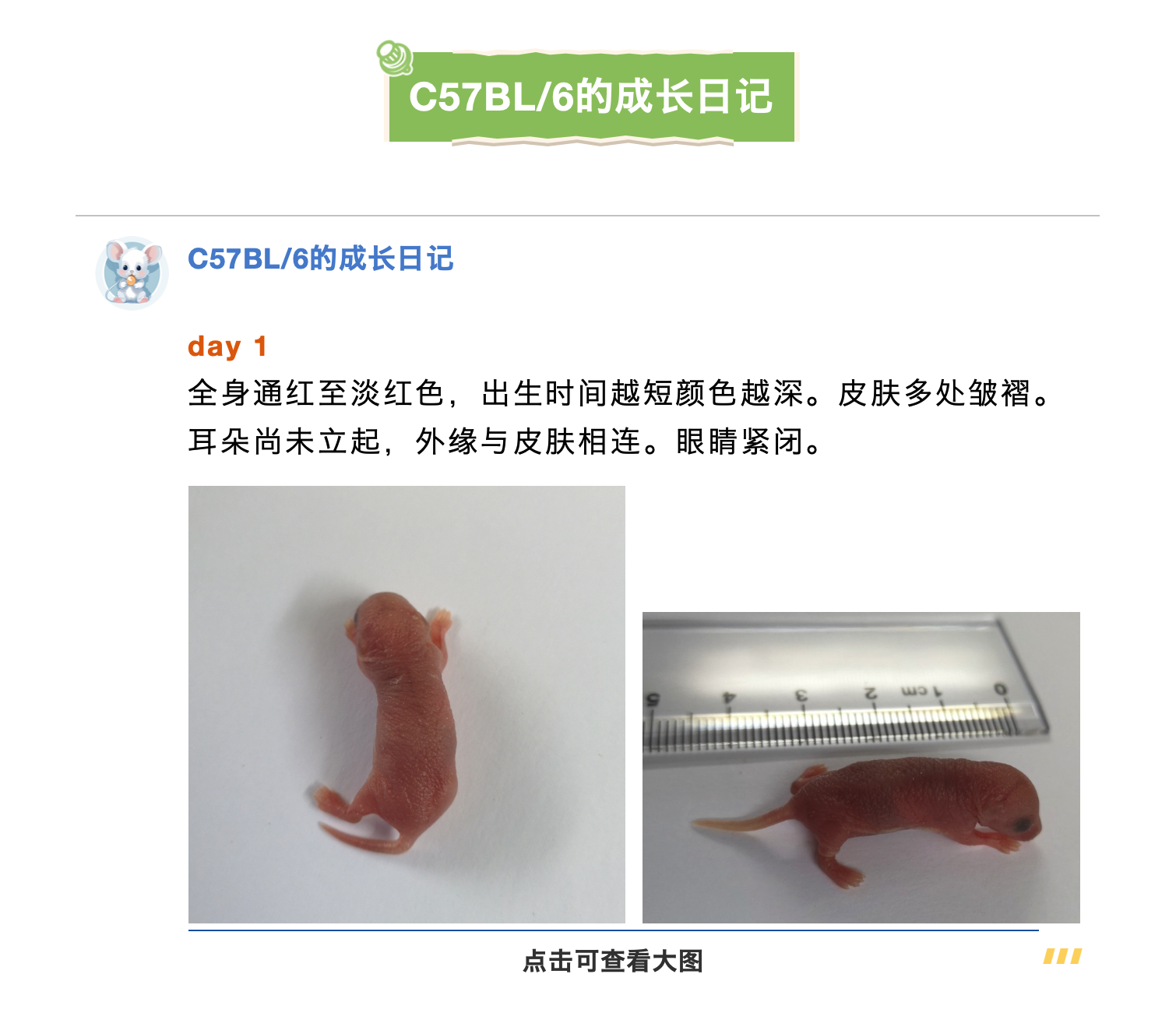
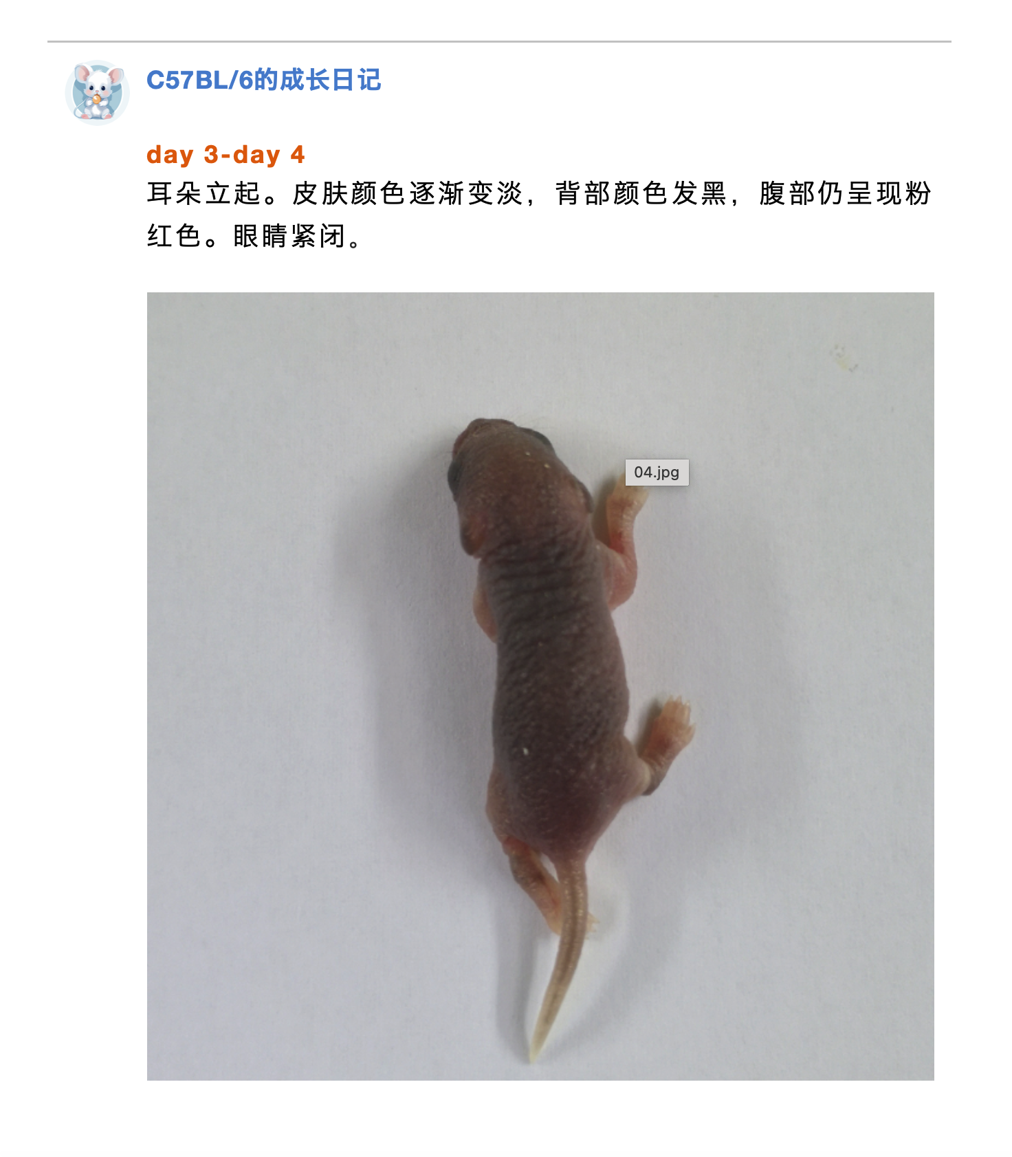


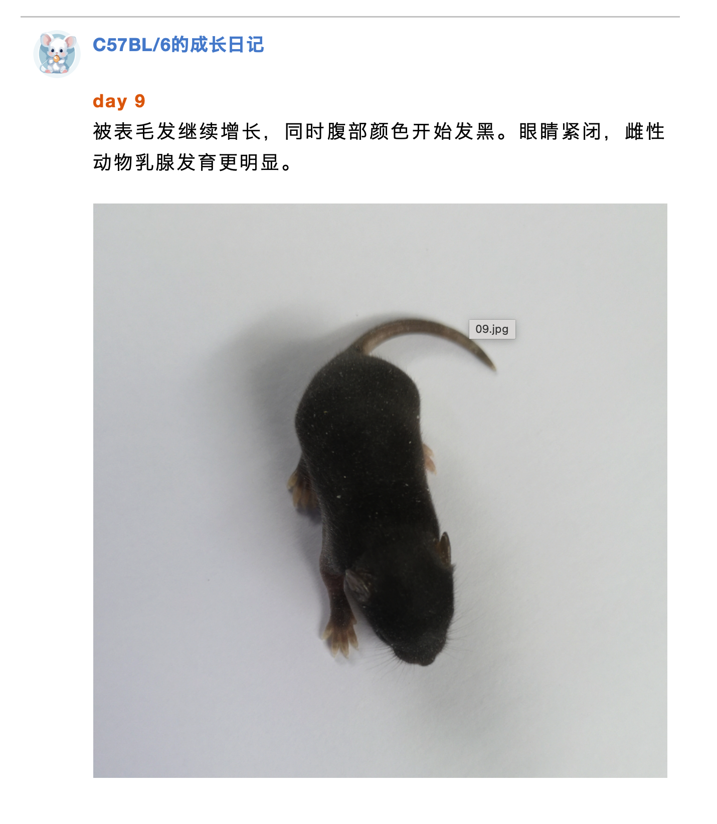
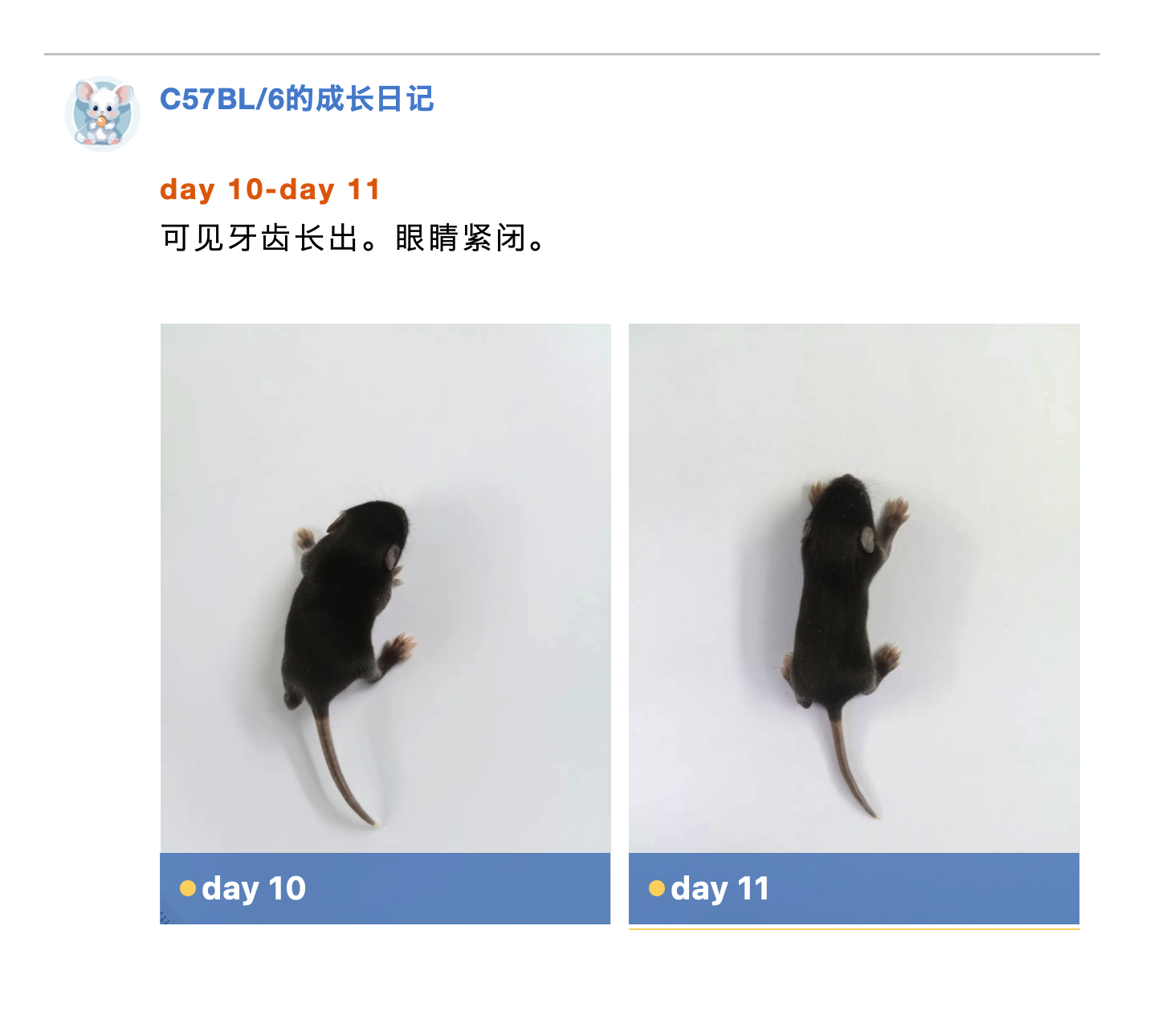
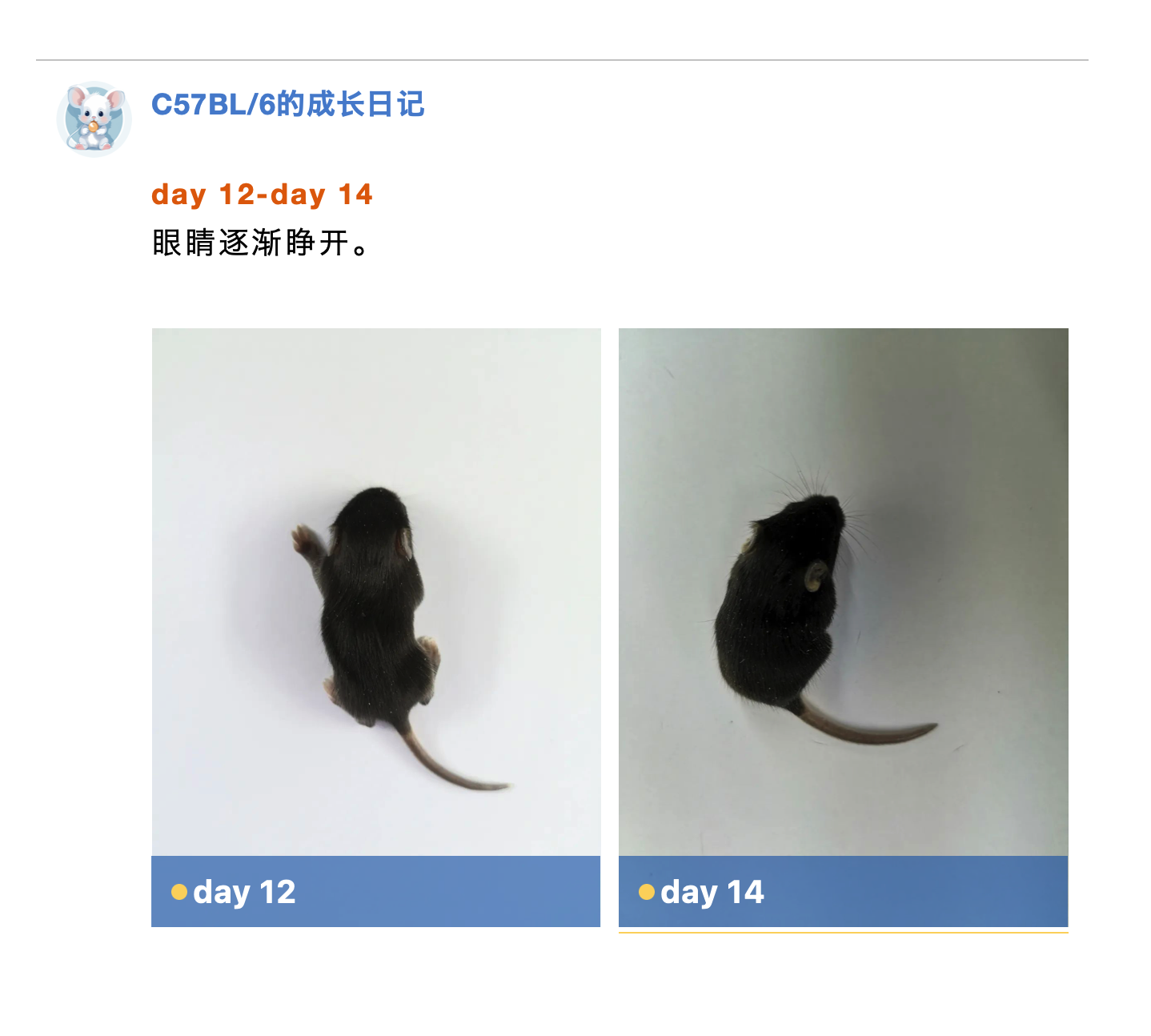
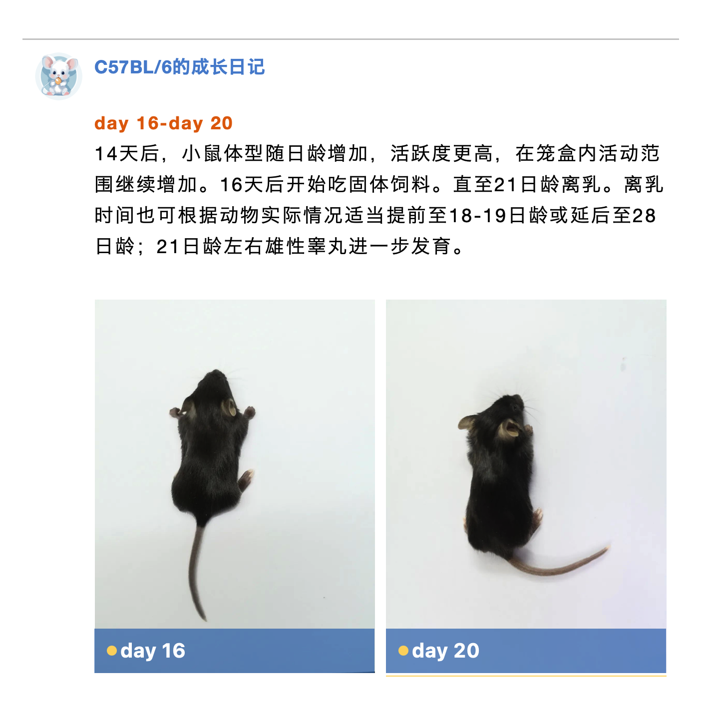
其他颜色小鼠(如BALB/c,DBA/2等)和大鼠(CD)在0-21日龄间发育变化特点与C57BL/6类似,可以通过几个重要变化来大致判断乳鼠的日龄范围:

裸鼠(Foxn1nu/Foxn1nu)的毛发受到Foxn1基因缺失的影响,绝大部分无法突出体表,在离乳前整体呈现全身光裸的状态,部分个体在头面部、颈部有稀疏毛发长出。同窝的杂合子个体(Foxn1nu/+)被毛变化与普通鼠无异。除毛发外,裸鼠及同窝杂合子动物的其他外观变化参考普通小鼠。


1. M. Hayward, Alison, et al. "The Mouse in Biomedical Research." (2007).
2. Adkins, B., Leclerc, C. & Marshall-Clarke, S. Neonatal adaptive immunity comes of age. Nat Rev Immunol 4, 553–564 (2004). https://doi.org/10.1038/nri1394
3. Di Bella, D.J., Habibi, E., Stickels, R.R. et al. Molecular logic of cellular diversification in the mouse cerebral cortex. Nature 595, 554–559 (2021). https://doi.org/10.1038/s41586-021-03670-5
4. Van Der Heijden, Meike E et al. “Quantification of Behavioral Deficits in Developing Mice With Dystonic Behaviors.” Dystonia vol. 1 (2022): 10494. doi:10.3389/dyst.2022.10494
5. Martin Aispuro, Pablo et al. “Use of a Neonatal-Mouse Model to Characterize Vaccines and Strategies for Overcoming the High Susceptibility and Severity of Pertussis in Early Life.” Frontiers in microbiology vol. 11 723. 17 Apr. 2020, doi:10.3389/fmicb.2020.00723
6. Philippot, Gaëtan et al. “Paracetamol (Acetaminophen) and its Effect on the Developing Mouse Brain.” Frontiers in toxicology vol. 4 867748. 22 Mar. 2022, doi:10.3389/ftox.2022.867748
7. McClain, R M et al. “Neonatal mouse model: review of methods and results.” Toxicologic pathology vol. 29 Suppl (2001): 128-37. doi:10.1080/019262301753178537
8. PIETRA, G., SPENCER, K. & SHUBIK, P. Response of Newly Born Mice to a Chemical Carcinogen. Nature 183, 1689 (1959). https://doi.org/10.1038/1831689a0
9. Sreejit, P et al. “An improved protocol for primary culture of cardiomyocyte from neonatal mice.” In vitro cellular & developmental biology. Animal vol. 44,3-4 (2008): 45-50. doi:10.1007/s11626-007-9079-4
10. Peter, Angela K et al. “Biology of the cardiac myocyte in heart disease.” Molecular biology of the cell vol. 27,14 (2016): 2149-60. doi:10.1091/mbc.E16-01-0038
11. Deeney, Scott et al. “A comparison of sexing methods in fetal mice.” Lab animal vol. 45,10 (2016): 380-4. doi:10.1038/laban.1105


